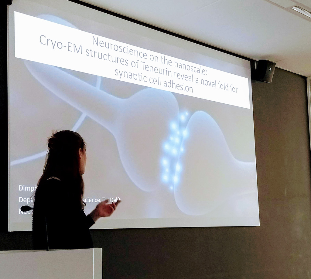
Abstract:
At the molecular level, synapse formation is enabled by cell-adhesion molecules that interact across the synapse. At present, many individual proteins have been identified that contribute to synaptic cell adhesion, but it is unclear how these individual proteins team up in macro-molecular complexes to organize synapse formation and function. We use single particle cryo-electron microscopy complemented with biophysical and cellular techniques to understand the molecular mechanisms of neural circuitry formation. Teneurins, a family of type II transmembrane proteins, have been characterized as synaptic cell adhesion molecules (CAMs) and are instrumental in the formation of neural circuits, including visual and memory circuitry. Previously, we determined the cryo-EM structure of Teneurin3 ectodomain in monomeric form. This reconstruction revealed a surprising novel fold for mammalian proteins. The structure contains a YD-shell topped with a beta-propeller similar to bacterial toxins, but it also harbors additional N- and C-terminal domains. Our current studies focus on the functional implication of the Teneurin domain architecture in developing neural circuits.

Video recording of the presentation to be uploaded at a later stage